
An amoeba is an organism unicellular highly motile eukaryote. It usually belongs to the kingdom of the protozoa and moves in an “amoeboid” manner. As such, microbiologists often use the term “amoeboid” to refer to a specific type of movement and amoebae interchangeably. Interestingly, amoebae are not a distinct taxonomic group and are instead characterised on the basis of their “amoeboid” movement rather than distinct morphological features. Moreover, even members of the same species may look different. Species of amoebae can be found in all major lineages eukaryotesincluding mushrooms, algae and even animals.
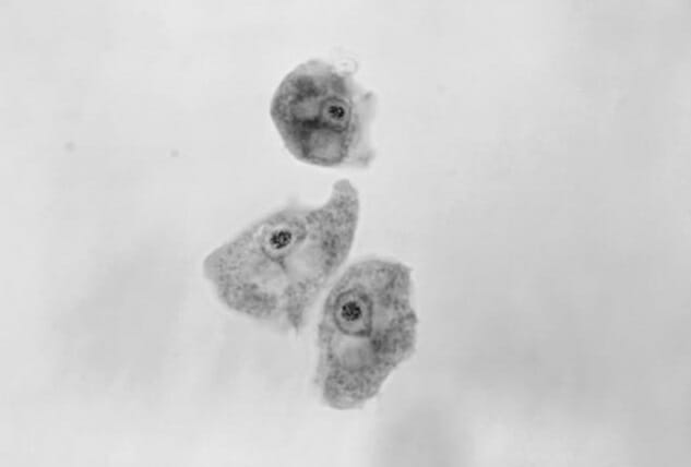
Amoebae contain an endoplasm that is granular in nature. This granular endoplasm contains the nucleus and several engulfed food vacuoles. In addition, amoebae are eukaryotes by definition and possess a single nucleus containing a central karyosome with a thin layer of bead-like chromatin that encases the nuclear membrane However, unlike many eukaryotes, amoebae are anaerobic. Therefore, amoebae do not contain mitochondria and generate ATP exclusively through anaerobic means.
Amoebae can be classified as free-living and parasitic. Parasitic amoebae are ubiquitous and often parasitise both vertebrates and invertebrates higher invertebrates. Only a limited number of amoeba species are capable of infecting humans and they usually invade the gut. Specifically, only Entamoebahistolytica represents a true human pathogen, which infects the gastrointestinal tract. A second intestinal pathogen, Dientamoeba fragilisis commonly mistaken for an amoeba due to its similar morphology under a light microscope. In fact, D. fragiliswas originally misclassified as an amoeba; however, modern methods have identified it as a non-flagellated trichomonad parasite. Interestingly, some free-living amoebae can cause opportunistic infections in humans, leading to eye infections, as well as various neurological and cutaneous infections (from the skin).
Amoeba movement
As a class of organism, amoebae are defined by their unique movement patterns. This movement strategy produces forward movement through the following three steps:
- “Swelling” the plasma membrane forward. This distinctive rearrangement is known as a pseudopod or “foot false”, which is very similar in nature to the lamellipodium generated in higher vertebrates;
- the pseudopod attaches itself to the substrate and is filled with cytosol;
- the back of the amoeba releases its attachment to the substrate and propels itself forward.
During amoeboid movement, the viscosity of the cytosol circulates between a fluid-like sol, which flows from the central region of the cytoplasm known as the endoplasm towards the pseudopod at the front of the cell. Once this occurs, the endoplasm becomes an ectoplasm containing a gel-like substance that forms the crust beneath the plasma membrane. As the amoeba moves on, the ectoplasmic gel once again becomes the endoplasmic sol, and the cycle repeats as the cell continues to move.
This transition between the gel and sun states occurs after the collapse and reassembly of the actin microfilament networks located in the cytosol. In particular, cofinin is responsible for the disassembly of actin filaments to form the sol, while profilin leads to actin polymerisation and the gel is formed by α-actinin and filamin.
Size and shape of the amoeba
Size
Amoebae differ in both size and shape, and even members of the same species can be very different morphologically. Although the first amoebae identified were approximately 400-600 microns in size, extremely small (2-3 microns) and exceptionally large (20 cm; visible to the naked eye) amoebae have been documented to date. Thus, amoeba species exhibit a wide range of sizes. In fact, when scientists study amoebae, samples are usually passed through a filter approximately 0.45 microns in size and the remains of the filter are used for culture.
Shape
Since amoebae move and eat using pseudopods, they are classified according to the morphology and internal structure of their pseudopods. For example, species of amoebozoans (e.g., amoebozoa) are classified according to their pseudopod morphology and internal structure, Amoebae ) exhibit bulbous pseudopods with a tubular midsection and rounded ends; Cercozoan amoeboids (e.g., cercozoan amoeboids) exhibit bulbous pseudopods with a tubular midsection and rounded ends, Euglypha y Gromia ) have pseudopods that appear slender and filiform; Foraminifera produce slender pseudopods that branch and fuse together to form net-like structures; others are characterised by rigid, needle-like pseudopods with a complex network of microtubules.
Free-living amoebae (which do not require a host) are either “testate” or “naked”. Testate amoebae contain a hard shell, whereas naked amoebae do not. The shells of testate amoebae are typically composed of calciumsilica, chitin or other components (e.g. sand granules).
Another component that is typically found in freshwater amoebae is a vacuole contractile. This vacuole is necessary to expel excess water from the cell and maintain osmotic balance. Since the concentration of solutes in fresh water is lower than in the internal cytosol of the amoeba, water flows through the cell membrane through the osmosis. Therefore, the contractile vacuole pumps this excess water out of the cell to ensure that the cell does not burst. In contrast, most marine amoebae do not have a contractile vacuole, as the cytosol and water outside the amoeba are balanced.
Reproduction of amoebae
Due to the extremely diverse nature of amoebae, the various species of amoebae reproduce using a variety of different methods. These methods include spores, binary fission and even sexually.
Binary fission
By far the most common form of asexual reproduction employed by amoebae is binary fission. In preparation for reproduction, the amoeba will remove its pseudopods and form a spherical shape. It is observed mitosis in the nucleus and the cytoplasm divides in the centre of the cell and separates, forming two daughter cells. Since this process involves simply copying the information genetic to form a second cell, the two resulting daughter cells are identical clones of the parent cell. The nucleus is therefore absolutely essential for this form of reproduction. This has been verified in experiments involving cutting an amoeba in half or removing the nucleus from the amoeba. In both situations, the cell eventually dies without a nucleus.
Multiple fission and encystment
Under conditions of food scarcity, amoebae will reproduce by multiple fission. This process involves the production of multiple daughter cells by:
- the pseudopods retract and the amoeba forms a spherical shape;
- the amoeba secretes a substance that hardens and encapsulates the cell, forming a cyst (encystment);
- the amoeba, protected by the cyst, will undergo mitosis several times, producing multiple daughter cells;
- when favourable conditions return, the cyst wall bursts, releasing the daughter cells. Within a host, the amoeba will undergo encystation as a means of protection against desiccation as it travels through the colon, ensuring its survival outside the host.
Spore formation
Solitary haploid amoebae (known as myxamoebae or “social amoebae”) reside in decaying vegetation (e.g., logs), eat bacteria, and reproduce asexually through binary fission as described above. However, unlike amoebae, which undergo encystment when food supply is depleted, tens of thousands of myxamoebae will fuse, forming a stream of moving cells that converge at a central location. It is in this region that the cells stack on top of each other and form a conical mound called a ‘compact aggregate’. A tip then rises from the top of the conical mound and the tightly packed aggregate folds to produce a mobile grex (also called a pseudoplasmodium or slug), 2 to 4 mm long and surrounded by a slimy substance.
The grex will then migrate to an illuminated area, where it will differentiate into a fruiting body composed of a tubular stalk (approximately 15% to 20% of the population total cell population) and spore cells. This process involves the secretion of an extracellular coating and the extension of a tube through the grex by the prestalk cells located in the anterior part of the grex. As the stalk cells differentiate into stalk cells, they create vacuoles and enlarge. This serves to raise the prespore cells in the posterior section of the grex. The raised prespore cells differentiate into spore cells and disperse, each representing a new myxamoeba, while the stalk cells die.
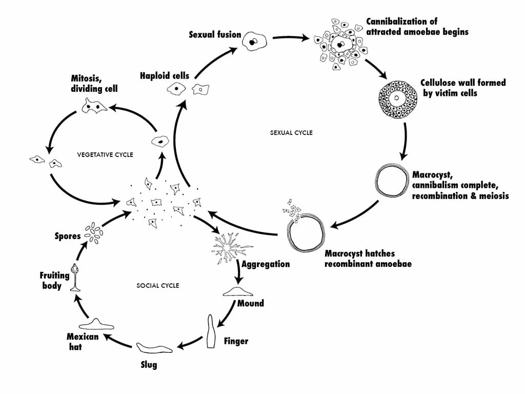
Sexual reproduction
Myxamoebae are also unique in that they can also reproduce sexually. This occurs when two myxamoebae fuse together to create one giant cell. This giant cell will then engulf all the other cells in an aggregate of myxamoebae. After engulfing all of its neighbours, the giant cell will become encysted and undergo division of the myxamoebae. meiosis and mitosis several times under the protective cover of the cyst. When the right environmental conditions are met, the cyst will burst and release new myxamoebae. Since this process involves meiosis and genetic information from two amoebae, the resulting daughter cells will be genetically distinct from the parent cells.
Temperature and reproduction
Temperature is a critical factor affecting the growth of amoebae. While various species of amoebae have been found to grow over a wide range of temperatures from 10°C to 37°C, pathogenic strains have been found to survive more efficiently at higher temperatures (between 32°C and 37°C). This indicates that amoebae are highly resistant to temperature fluctuations and most are adapted to survive inside humans. This may therefore have pathogenic implications, as amoeboid cysts are extremely resistant to microbicides and can infect humans through contaminated drinking water.