
Definition of centromere
The centromere is the point on a chromosome where the fibres of the mitotic spindle join together to separate the sister chromatids during the cell division.
When a cell seeks to reproduce itself, it must first make a full copy of each of its chromosomes, to ensure that its daughter cell receives a full complement of the DNA of the stem cell.
The two copies of each chromosome often remain attached until they separate, with one copy for each daughter cell. While stuck together, these two copies are called “sister chromatids”.
As a cell prepares to divide, the sister chromatids begin to detach from each other until they are almost completely separated. However, they remain attached at the centromere, a special region that plays a vital role in cell division.
At the centromere, the elements of the cytoskeleton of the cell assemble and attach. First, a protein complex called the kinetochore assembles around the centromeric region of the DNA; then, mitotic spindle fibres attach to the kinetochore. The other end of these fibres is anchored to opposite ends of the mother cell, which will shortly divide to become new daughter cells.
When the spindle fibres begin to contract, the chromatids are pushed towards opposite ends of the mother cell. Thus, when the stem cells divide in two during the cytokinesiseach chromatid sister becomes a chromosome of the new daughter cell.
To understand this process, it is important to remember that each sister chromatid is actually a complete copy of the parent cell’s chromosome.
The two sister chromatids combined are often referred to as a single chromosome because they are packed closely together, but each contains all the information from the original chromosome, so when they divide, each becomes a complete chromosome containing all the information contained in the cell’s original chromosome matrix.
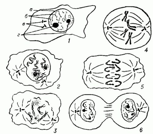
The image below provides a visual illustration of the cell preparations to undergo cell division. Note that in stage 2 the nuclear envelope dissolves, leaving the chromosomes in the cell free. cytoplasm.
In stages 3 and 4, the DNA condenses into compact chromosomes, in which the sister chromatids pair up and join at their centromere. In stage 5, shown below, the sister chromatids separate on opposite sides of the cell.
In stage 6, the cell finally splits in two, separating the sisters into daughter cells.
Role of the centromere
All living things are made up of cells. For cells to grow or reproduce, cell division must occur. In cell division, a “mother” cell divides into two, and each of the resulting cells are “daughter” cells.
For each daughter cell to survive, it is essential that they get a copy of the chromosomes of each of the parent cells.
When this does not happen and the daughter cells receive incomplete information, or too many copies of a chromosome, serious disease or cell death can result.
To ensure that each daughter cell is given a complete copy of its DNA, a cell first makes a complete copy of its DNA. The two copies stick together and eventually condense to form sister chromatids, until they separate during cell division.
The centromere of the chromosome provides a binding site for the mitotic spindle fibre that will attach to each sister chromatid and push them towards opposite ends of the parent cell, which will eventually become the cytoplasm of the two daughter cells.
In cases where centromeres do not function properly, cells cannot divide successfully. Any attempt to do so results in daughter cells that do not have the genetic instructions they need to survive.
Centromere dysfunction leading to problems with chromosome sorting is thought to play a role in many cases of miscarriage, where inherited centromere disorders can lead to early embryonic death. Centromere dysfunction is also suspected to play a role in cancer cells, which show massive chromosome imbalance of the type that would be expected if chromosome sorting fails during cell division.
Centromere types
Point centromeres
Point centromeres are centromeres where mitotic spindle fibres are attracted to specific DNA sequences. In these cases, the cell has proteins that bind to these specific DNA sequences, and these proteins form the basis for the binding of the mitotic spindle fibres.
In these cases, mitotic spindle fibres will typically appear wherever the DNA sequence of the punctate centromere appears. The protein that initiates the creation of the mitotic spindle fibre complex will bind to that DNA sequence regardless of its location or other factors.
Regional centromeres
Humans and most cells eukaryotes use regional centromeres. These are centromeres where the mitotic spindle junction is determined, not by a precise DNA sequence, but by a combination of features that work together to pinpoint the location of a centromere.
At regional centromeres, epigenetic marks are thought to tell proteins that begin to build the mitotic spindle complex where to bind.
Epigenetic marks” are chemical changes made to DNA by enzymes, which can change DNA’s chemical and other properties. Epigenetic marks can be added or removed without changing the information contained in the DNA.
- Epigenetic marks: chemical changes that enzymes can make to DNA. Epigenetic marks are reversible and are thought to play a role in the organisation of chromosome formation and the regulation of gene expression.
- Kinetochorea: protein structure that forms around centromeres during cell division. It is the kinetochore to which the mitotic spindle fibres attach.
- Mitosis: the most common type of cell division used by eukaryotic cells. Under specific circumstances, other methods of cell division may be used, such as meiosis.